


Digital images obtained entirely from systems operating with digital technology are stored in a digital archive. Digital images are more detailed and better for diagnosis compared to images obtained with conventional methods. Such images are analyzed by specialists in dedicated rooms. In our Radiology department, radiological examinations such as the following are performed:
Today, magnetic resonance imaging is used for imaging soft tissues. It is frequently used in diagnosing central nervous system diseases (brain and spinal cord), sports injuries, musculoskeletal system, especially in detecting disorders such as meniscus and herniation, and in evaluating all types of neurological diseases. To date, no proven harm from magnetic resonance imaging to living organisms has been established, including in pregnant women. However, MRI is not recommended during the first three months of pregnancy, when the baby's organs are developing.
Since it can be life-threatening, it is considered unacceptable for persons who have metal-reactive substances in their body, such as magnets, metal prostheses, pacemakers, foreign bodies in the eye, or persons who have suffered gunshot wounds, to enter the MRI machine.
The duration of an MRI scan varies depending on the number of areas to be examined and the preliminary diagnosis. The duration can range from 15 minutes to 75 minutes.
The X-ray beam rotates 360 degrees around the patient, passes through detectors arranged in a cavity that detect the portion of X-rays passing through the body, and the obtained data is processed in a computer. As a result, cross-sectional images of the tissues are formed, which can be viewed on a computer.
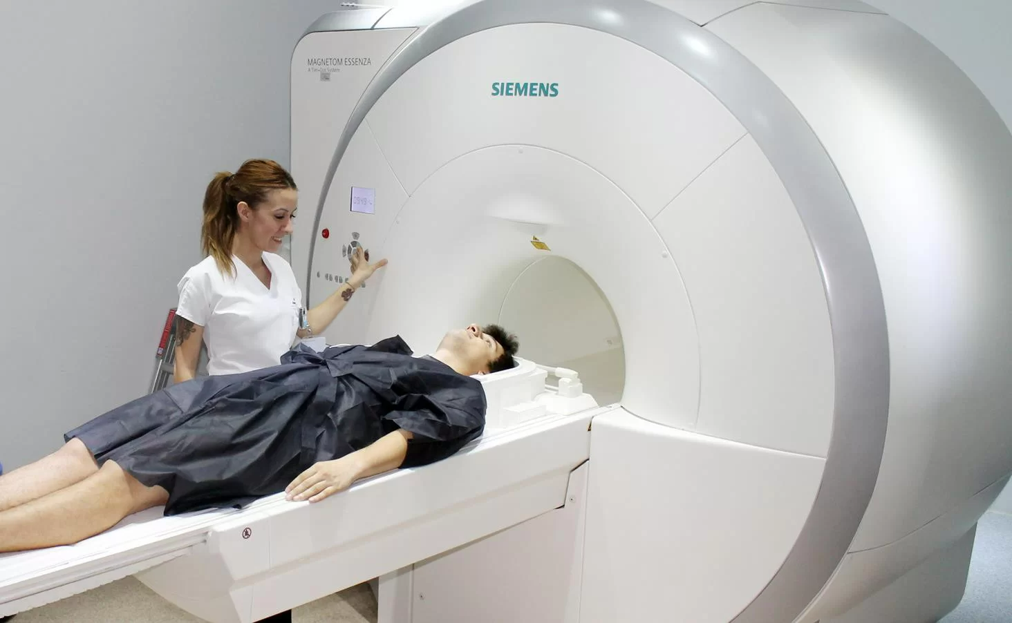
The technician takes the patient to the room where the procedure will be performed. Depending on the examination to be performed, they instruct the patient to lie on the table on their back or stomach. It is important for the patient to remain calm, as they must stay still during the examination.
Tomographic examinations vary depending on the medical problem and the part of the body to be scanned. The staff working in this field holds the necessary certifications and is well-trained to provide the best services for patients.
From the perspective of breast cancer diagnosis, mammography is an important examination method for detecting minimal abnormalities in the breast that cannot be identified during a normal examination.
Mammography can be performed on any woman from a medical standpoint, but it is recommended to be done at least once a year for patients between the ages of 35-40.
During imaging, the breast is compressed between the two sides of the device for a few seconds. For this reason, it is recommended that mammography be performed at a time when the breasts are least sensitive, especially for women with sensitive breasts.
 Phone
Phone