Last updated: 17 февруари 2026
To schedule an appointment, leave us your contact information and our English-speaking colleagues will contact you to provide more detailed information.
Videos

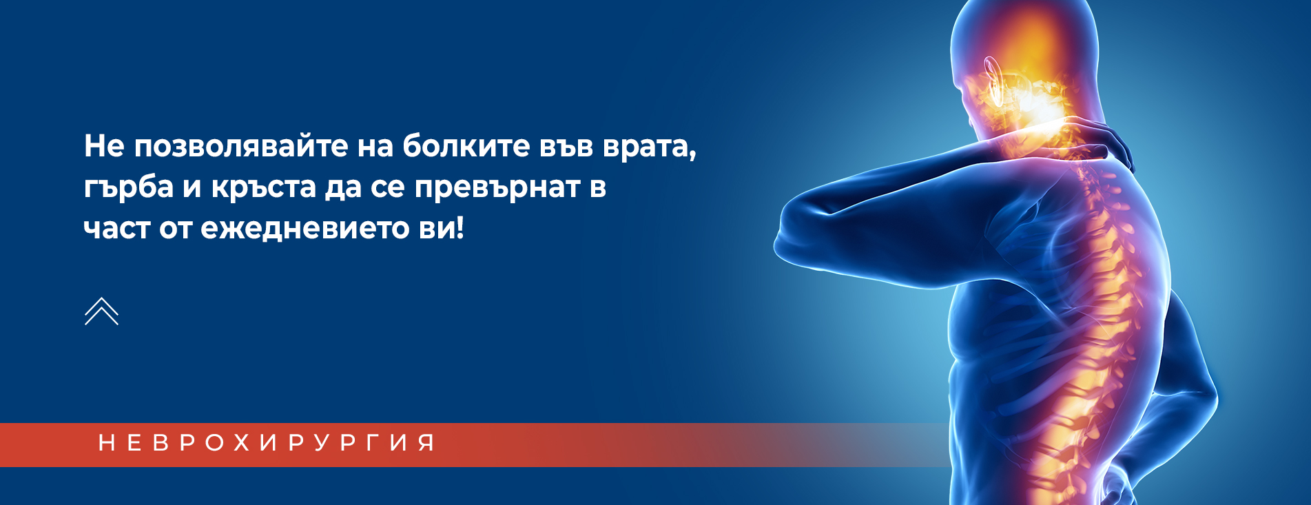
At the Neurosurgery Department of Corlu Vatan Private Hospital, we offer precise examination, diagnosis, and modern surgical treatment of diseases of the central and peripheral nervous system.

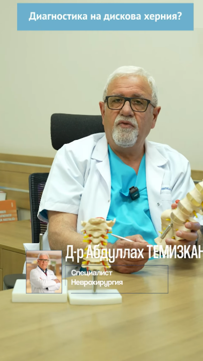
The diagnostic process begins with a thorough physical examination by a Neurosurgery specialist. It consists of palpation of painful areas, checking tendon reflexes, skin sensitivity, muscle strength, and signs of nerve root tension. After the physical examination, the experienced specialist can easily identify the potential cause of the patient's complaints.

In this field of medicine, a physical examination alone is not sufficient for making a definitive diagnosis. The diagnosis is established after additional imaging studies such as: X-ray, Computed Tomography (CT), and Magnetic Resonance Imaging (MRI).

  • Using X-rays in examinations such as X-ray imaging and computed tomography, specialists can gain a clear understanding primarily of the patient's skeletal system. The significance of these examinations is more indicative in diseases such as osteoporosis, inflammatory diseases, injuries, bone tumors, etc.
  • MRI scanning is a non-invasive examination using a strong magnetic field and radio frequency waves to provide a detailed view of the soft tissues (nerves and intervertebral discs) of the respective area being examined. Today, MRI is accepted as the gold standard in diagnosing spinal diseases. With the help of MRI, it is possible to determine which intervertebral disc is damaged and whether there is any compression on the nerve roots. The examination is also used in diagnosing brain tumors, spinal cord tumors, and many other diseases.

By applying the most advanced achievements of medicine and providing first-class healthcare comparable to world standards, we strive to offer our patients appropriate treatment tailored to their needs and improvement of their quality of life.

Frequently Asked Questions
  • Treatment of Spinal Stenosis (narrowing of the spinal canal)

    Spinal canal stenosis (spinal stenosis) is the narrowing of the space at one or more locations along the spinal canal. A narrowed spinal canal can lead to compression of the nerve structures (spinal cord and nerve roots) that pass through the spinal column. Spinal stenosis is most commonly found in the lumbar and cervical regions. Some patients with spinal stenosis may have no complaints, while another group of patients may experience symptoms such as pain, numbness, decreased sensation, and muscle weakness. These symptoms may intensify and worsen over time.

    Treatment of spinal stenosis varies depending on the severity of symptoms and the exact location of the stenotic segment. Lack of response to conservative treatment and the presence of persistent pain or progressive neurological deficit and pelvic reservoir disorders necessitate surgical intervention.

    The choice of surgical technique is of exceptional importance, as the type and extent of the surgical intervention are determined by the nature of the stenosis. Decompression of the affected nerve elements is necessary without causing spinal instability. Removal of hypertrophied bony segments requires exceptional care to avoid causing additional damage to neural structures in the narrowed space.

    Implant-free treatment of spinal stenosis using the microsurgical method!

    This method aims to perform decompression in the respective area of spinal canal narrowing without placing foreign bodies (implants) in the patient's body. With this method, the narrowed canal is widened from the inside out. This is one of the most modern methods for treating spinal stenosis and has numerous advantages compared to classical surgical techniques. Some of these advantages are:

    • The surgical intervention is much shorter in duration compared to classical open surgery.
    • The patient's postoperative treatment period is significantly alleviated, and therefore the recovery period is much shorter.
    • The patient can return to their normal daily life and work in a much shorter time.
    • Due to the fact that no implants are used, the risk of potential postoperative infection is minimized.
    • The risk of complications such as paralysis, nerve damage, etc. is much lower compared to classical open surgery.

    For the above-mentioned reasons, implant-free treatment of spinal stenosis using the microsurgical method is also preferred for elderly patients with accompanying conditions.

  • Treatment of Spondylolisthesis (vertebral slippage)

    Spondylolisthesis is the displacement (slippage) of one vertebral body of the spinal column relative to another. The displacement can occur in all directions - forward, backward, or sideways, and can lead to compression of nerve roots and narrowing of the spinal canal. This can lead to compression of the spinal cord and serious damage to some neurological functions.

    Spondylolisthesis most commonly affects the lower part of the lumbar region of the spinal column. It can manifest at all ages. The initial stages of the disease may progress without symptoms for varying periods of time. In the advanced stages of the disease, as compression on the nerve roots and/or spinal cord increases, pain gradually begins in the area of slippage along with neurological complaints. The pain and sensations may spread to the lower extremities and can cause muscle weakness in the limbs, muscle spasms, numbness, and tingling along the path of the affected nerves.

    The main causes of spondylolisthesis are congenital vertebral displacement, trauma from accidents or sports activities, and degenerative changes of the spinal column. The most commonly encountered form of the disease is degenerative spondylolisthesis. It is the result of changes that occur in the vertebrae during the aging process.

    TREATMENT OF SPONDYLOLISTHESIS

    With advancing age, the vertebrae begin to lose their stability, dehydration occurs, which in turn leads to their cracking. This creates favorable conditions for the displacement of vertebrae from their original position.

    In mild forms of the disease, treatment is primarily carried out through physiotherapy and rehabilitation, balneotherapy, and strengthening of the muscles of the affected segment of the spinal column. Patients are also prescribed pain-relieving medications, as well as vitamins and minerals to strengthen the skeletal system. Wearing a special supportive medical corset also alleviates the patient's condition.

    In more severe forms of the disease, vertebral displacement leads to increasing pain symptoms and the possible appearance of neurological deficit. This in turn leads to serious risks of permanent damage and necessitates surgical treatment.

    Surgical treatment of spondylolisthesis includes decompression of nerve structures, restoration of normal anatomical relationships between vertebrae, and stabilization for the purpose of fusion. In surgical treatment, classical operative techniques for decompression are combined with modern instrumental stabilization, with spinal implants placed to achieve spinal stability.

    The goal of the surgical intervention is to achieve complete surgical decompression of neural structures and maximum stability of the damaged segment of the spinal column.

  • Treatment of Brain Tumors

    Every living organism is composed of numerous cells of different types. Sometimes, under the influence of various and very often unclear causes, some of our cells change their normal development cycle. These cells begin to divide abnormally and uncontrollably, thus forming a new growth - a tumor. When this new growth develops in the brain, it is identified as a brain tumor.

    There are various types of brain tumors. Depending on the classification criteria, brain tumors are classified in several different ways.

    The majority of brain tumors are formed from cells of the brain tissue itself. Brain tumors that originate from brain tissue, brain membranes, and cranial nerves are classified as primary brain tumors. Secondary brain tumors, also called metastatic brain tumors, originate from organs and tissues outside the brain (when an oncological disease in another part of the body spreads to the brain) and reach it through the bloodstream.

    Depending on the differentiation of the cells and tissues from which they are composed, brain tumors are classified as benign and malignant.

    TREATMENT OF BRAIN TUMORS

    Treatment of brain tumors is a complex process and often requires the involvement of a team of specialists. The treatment method is determined depending on the exact location, size, and type of the tumor and other individual characteristics of the patient.

    Surgical removal of benign brain tumors is a reliable treatment method with a high success rate. If the patient's case allows complete removal of the benign tumor during the surgical intervention, the problem will be resolved without the need for additional treatments. This procedure is a surgical procedure with a very high success rate and provides the opportunity for full recovery.

    Unlike other tumors in the body, brain tumors are distinguished by one important characteristic feature - they develop in the cranial cavity, which represents a closed space with rigid boundaries. For this reason, it is accepted that all brain tumors are relatively malignant, since the human brain has nowhere to "retreat" when compressed by the tumor. Another important characteristic of brain tumors is that even the presence of a small tumor located in a functionally significant area of the brain (motor centers, sensory zones, the speech center of the brain, etc.) can lead to serious damage and significantly worsen the patient's quality of life.

    Treatment of malignant brain tumors often requires comprehensive treatment encompassing neurosurgical operation, radiation therapy, chemotherapy, and rehabilitation. If the brain tumor is prognosticated as malignant, it is of primary importance to determine the exact type of tumor for establishing the treatment plan. In this way, planning of the patient's entire treatment can begin. For this purpose, a biopsy is performed (taking a small part of the tumor through a small opening in the skull for detailed analysis).

    If there are already serious negative effects on the patient's overall health condition, treatment procedures can begin directly with surgical interventions aimed at minimizing the negative effects of the brain tumor. Sometimes, due to the location of the brain tumor, it is not possible to remove it entirely during the surgical intervention. In such cases, subsequent treatment methods such as radiation therapy and chemotherapy are necessarily applied.

    Performing surgical intervention in this area of the human body requires the use of microsurgical methods. Due to the presence of numerous fine nerve formations in this area of our body, even the smallest problem can have major consequences. Therefore, surgical intervention on brain tumors is performed with the aid of neuronavigation (neuromonitoring). Through this system, the neurosurgeon monitors the patient via signals from electrodes placed on the patient's face, arms, and legs. When signals decrease, the doctor receives a warning that they are in a sensitive area of the brain. Thanks to this system, the risk of paralysis and unwanted negative consequences from the surgical intervention are minimized.

    The success of the surgical operation is of determining importance for the overall treatment of brain tumors.

  • Treatment of Disc Herniation

    WHAT IS DISC HERNIATION?

    The vertebrae forming the spinal column are arranged one on top of another and are connected to each other, forming the spinal canal. The spinal column protects the spinal cord from external influences. In addition to vertebrae, the spinal column contains the spinal cord, nerve endings, and cartilaginous tissue discs that provide flexibility to the spinal column. The outer wall of each of these round discs, located between the vertebrae, consists of elastic and firm tissue. The inner part of this cartilaginous tissue (disc) has a gelatinous structure. As a result of wear or injury to the outer wall of the cartilaginous disc, the gelatinous material located in the center begins to press the outer wall from the inside out. If the disc is very worn or severely damaged, the gelatinous material prolapses outward, thus forming disc herniations. The more commonly encountered types of disc herniation are lumbar and cervical disc herniation.

    DIAGNOSIS OF DISC HERNIATION

    A number of imaging studies are used in diagnosing disc herniation. Nowadays, Magnetic Resonance Imaging (MRI) is accepted as the most modern method and the gold standard for diagnosing spinal column diseases. After a physical examination and MRI, the specialist will be able to inform you in more detail about your current condition and the treatment necessary in your case.

    TREATMENT OF DISC HERNIATION

    Most patients do not visit a doctor because they fear that surgical intervention will be recommended, but only 10% of patients with lumbar and cervical disc herniations require surgical intervention, i.e., the remaining 90% can be treated non-surgically. The treatment plan for lumbar and cervical disc herniations is determined based on the patient's complaints, clinical condition, and imaging diagnostic results. Excluding emergency conditions, the first step in treatment is conservative therapy. If necessary, treatment continues using invasive surgery methods - trigger point injections, epidural injections, spinal anesthesia, and intradiscal manipulations. Surgical treatment is resorted to when conservative therapy is not sufficiently effective or in cases of muscle strength loss, urinary incontinence, and severe pain unresponsive to medication.

    In our Neurosurgery clinic, surgical interventions for disc herniation treatment are performed primarily using the microsurgical method. Before the surgical intervention, based on the physical examination and MRI, a decision is made regarding the most appropriate surgical method for the specific case.

    One of the most commonly applied modern treatment methods in this area of medicine is "Microscopic Discectomy." The term "Microdiscectomy" refers to the removal of the herniated part of the intervertebral disc through a minimal skin incision, using a surgical microscope under high magnification and with the help of special microsurgical instruments. The fundamental principles of microsurgery are preserving the maximum anatomical integrity of tissues. The operative incision is without cosmetic defect.

    The microsurgical approach has numerous advantages over standard surgical intervention:

    ➤ Significantly reduced operative time and minimal incision.

    ➤ Fewer risks of postoperative complications to musculature and nerves.

    ➤ Possibility of easy application in patients being treated for osteoporosis and/or diabetes.

    ➤ Fewer risks of postoperative infections.

    ➤ Significantly reduced hospital stay.

    ➤ Short recovery time and quick return to normal daily life.

Specialists in This Field

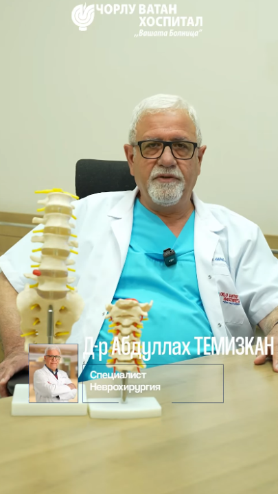
Dr. Abdullah TEMİZKAN graduated from the Faculty of Medicine at Dicle University. In 1989, he completed his specialization in Neurosurgery at the Faculty of Medicine at Cumhuriyet University. The specialist has many years of professional experience in the field of Neurosurgery in various regions of the Republic of Turkey. Since 2016, he has held an active position at Çorlu Vatan Private Hospital.

Dr. Oğuz ÖZEL graduated from the Faculty of Medicine at Trakya University. In 2011, he completed his specialization in Neurosurgery at the Faculty of Medicine at Erciyes University. The specialist has many years of professional experience in the field of Neurosurgery in various regions of the Republic of Turkey. Since 2020, he has held an active position at Çorlu Vatan Private Hospital.

Our Patient Stories
Yılan Yapısı 1555788326
Successful Treatment of a Fractured Cervical Vertebra After a Severe Traffic Accident

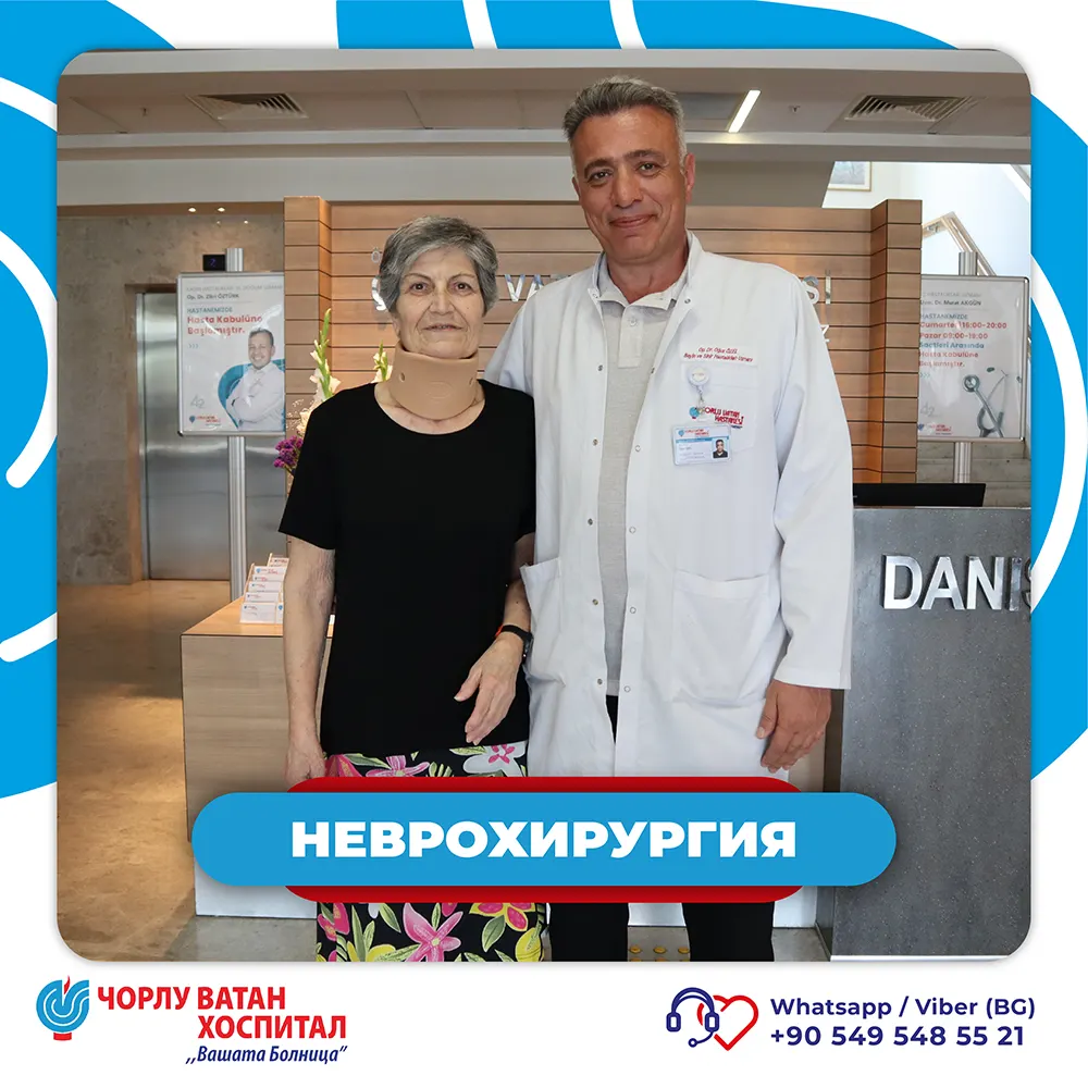
At Corlu Vatan Private Hospital, neurosurgery specialist Dr. Oguz YOZEL performed anterior odontoid fixation on a 19-year-old patient who suffered a severe traffic accident. The patient was referred to our hospital from another medical facility, and upon arrival, detailed radiological examinations were performed, which revealed a fracture of the second cervical vertebra (C2), which is crucial for neck movement and stability.

After careful evaluation of his condition, Dr. Oguz YOZEL performed anterior odontoid fracture fixation, which restored the stability of the spinal column. The surgery was successful, and just four days later the patient was discharged in good and stable overall condition.

Upon leaving the hospital, the patient expressed sincere gratitude to Dr. Oguz Yozel and the entire team of Corlu Vatan Hospital for their high professionalism, attention, and care provided throughout the entire treatment period.

The hospital team joyfully extends its warmest wishes for a speedy and complete recovery, as well as a long, happy, and healthy life for our patient!

Yılan Yapısı 1669817051
Treatment of Lumbar Disc Herniation and Spinal Stenosis!

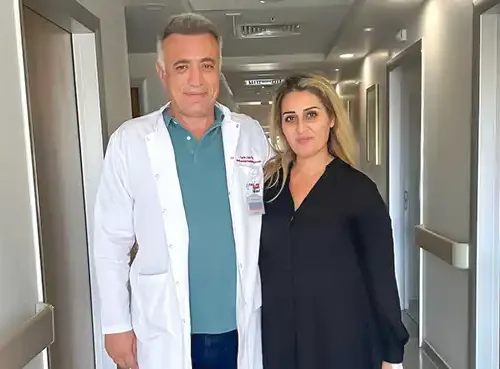
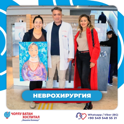
Our patient from Australia arrived at our hospital with complaints of lower back pain, numbness, and pain along the left leg. She shared with us that she had undergone two previous surgical interventions for disc herniation treatment.

The patient's case was taken on by Dr. Oguz YOZEL - neurosurgery specialist. After a thorough physical examination and imaging studies, it was determined that the patient's condition required a new surgical intervention.

The patient was offered the surgical procedure "Microdiscectomy and Foraminotomy," aimed at removing the herniated portion of the intervertebral disc causing pressure on the nerve roots, and widening the spinal canal in the corresponding segment of the spinal column.
One day after the successful surgical intervention, our patient was discharged in good health, having said goodbye to the complaints she had upon admission to the hospital.
She expressed her gratitude to Dr. Oguz YOZEL and the entire team that cared for her during her stay at our hospital.
The entire team of Corlu Vatan Private Hospital wishes our patient a speedy recovery, and a long and healthy life.

If you too suffer from lower back or leg pain, experience loss of sensation and numbness in your extremities, or wish for a specialist consultation, schedule an appointment with our specialists today!

Yılan Yapısı 1499018454
Treatment of Cervical Disc Herniation

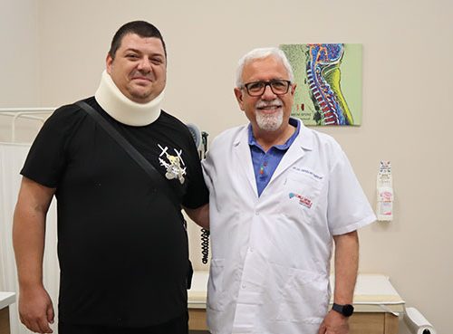
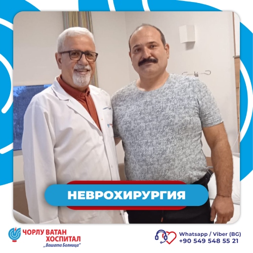
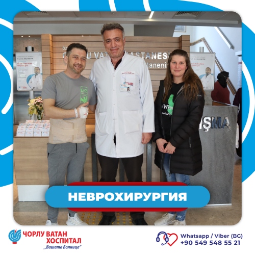
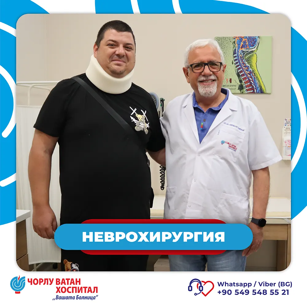
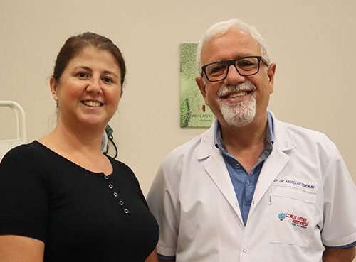
33-year-old Mr. Kristian Tonev turned to Corlu Vatan Private Hospital seeking relief from severe pain in his neck and left arm. After these pains began to significantly disrupt his daily life and overall well-being, he contacted the International Patients Department of Corlu Vatan Private Hospital to find a solution to his problem. Mr. Tonev's case was taken on by Dr. Abdullah TEMIZKAN - Neurosurgery Specialist.

After our patient arrived at the hospital, a detailed physical examination and imaging studies such as MRI were performed.

Based on the physical examination and imaging studies, the patient was recommended a surgical intervention to remove cervical disc herniation at two levels in the cervical spine. The patient accepted the proposed surgical intervention and the preoperative preparation phase began. After it was established that there was no medical obstacle to performing the surgical intervention, the patient was hospitalized.

The next day, the surgical intervention was successfully performed by Dr. Abdullah Temizkan, using a microsurgical method with an anterior approach. The microsurgical method offers a number of potential benefits for the patient, including reduced blood loss, faster recovery and return to normal daily life, and minimization of the risk of postoperative complications.

After a three-day recovery period and postoperative hospitalized observation, Mr. Tonev was ready to return home to Bulgaria, in improved health, having said goodbye to the pain he had upon admission to the hospital.

Happy with the successful outcome of his treatment, Mr. Kristian Tonev expressed his heartfelt gratitude to Dr. Abdullah TEMIZKAN and the entire team of Corlu Vatan Private Hospital for their care and professionalism.

Mr. Kristian Tonev is just one of many examples of successful treatments that we achieve daily at Corlu Vatan Private Hospital. We thank all our patients for their trust and are glad that we can contribute to their well-being.

Yılan Yapısı 234063268
VERTEBROPLASTY: A Successful Treatment Method for Patients with Osteoporotic Fractures

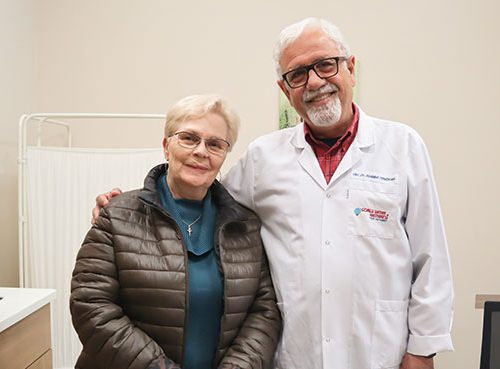
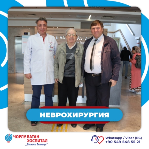
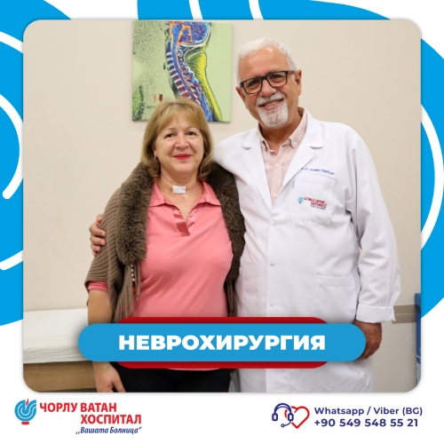
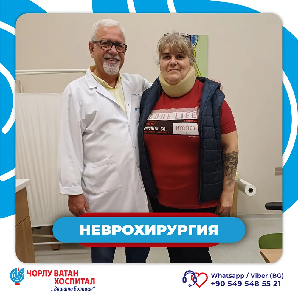
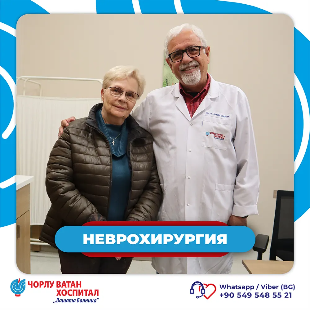
We would like to share with you the story and successful treatment of our 71-year-old patient who visited us from the Republic of Bulgaria.
By contacting our International Patients Department, Mrs. Tonka Boeva shared her complaints of severe pain in the back and lower back area. After analyzing the old examination results sent by our patient, she was invited for a physical examination and new tests to evaluate her current condition.

Upon her arrival, our patient was examined by our neurosurgery specialist Dr. Abdullah TEMIZKAN. After a detailed physical examination and analysis of the new radiological studies (MRI and CT Scan), several osteoporotic fractures were found in the thoracic and lumbar segments of the spinal column. Due to these fractures, the patient had assumed a kyphotic (hunched) posture.

Based on the patient's age, existing chronic conditions, and current health status, the Vertebroplasty intervention was proposed. The surgical intervention was performed the following day by our neurosurgeon Dr. Abdullah TEMIZKAN. To correct the acquired kyphotic posture (hunching), during the surgical intervention (using a kyphoplasty balloon), the bones were restored to their normal position and height.

During the intervention, Dr. Abdullah TEMIZKAN, under X-ray guidance, precisely filled the existing fractures and collapses by injecting a substance called bone cement. After two days of postoperative monitoring, our patient was discharged from the hospital in good health and with satisfaction from the achieved relief. She expressed her sincere gratitude to Dr. Abdullah TEMIZKAN and the entire team for their professionalism and the care they provided.

The entire team of Corlu Vatan Private Hospital wishes to express our most sincere wishes for a successful recovery of our patient. We wish her a speedy recovery and good health going forward!

Yılan Yapısı 893471571
Successful Treatment of Long-Standing Intensifying Pain!

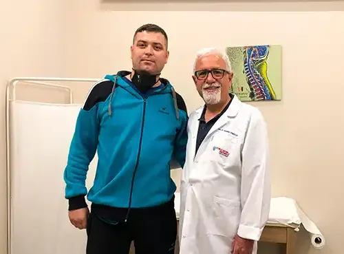
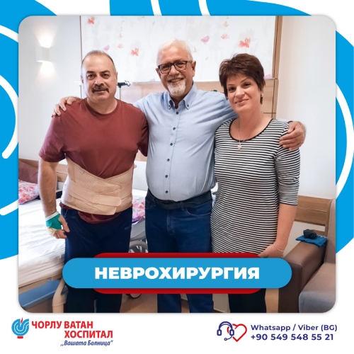
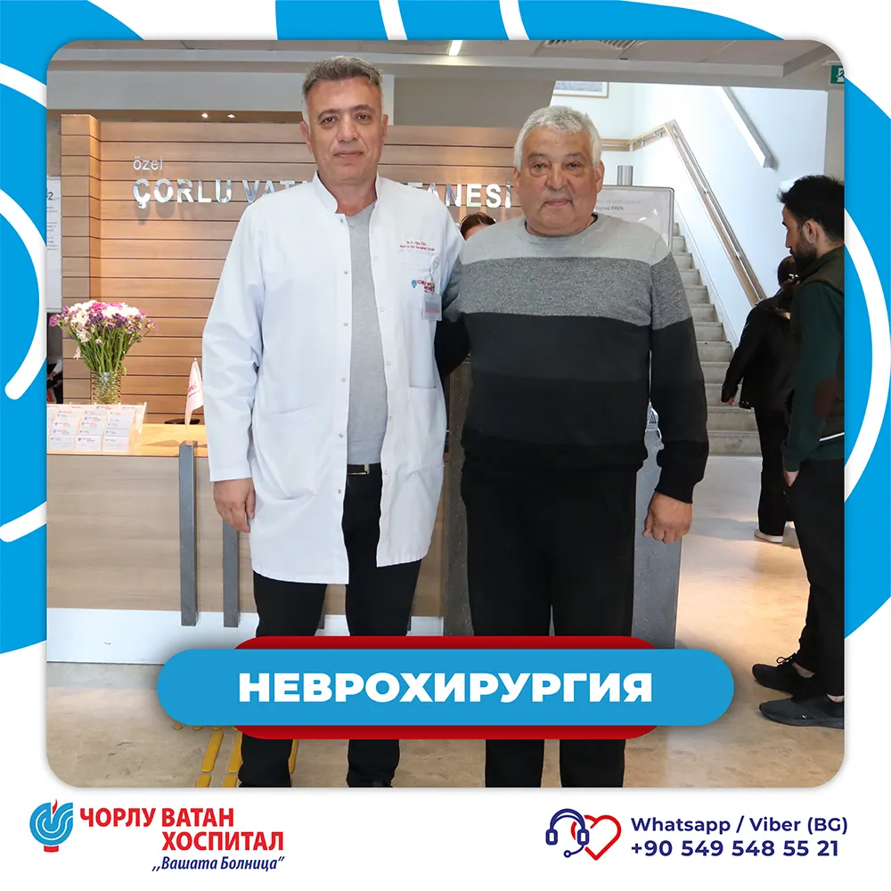
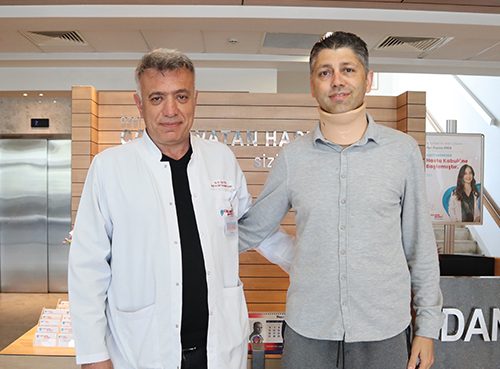
Our patient Mr. Yumer Beyti Ahmed from the city of Tervel, Bulgaria, contacted our International Patients Department requesting treatment for his complaints, which were in the field of Neurosurgery. He shared with us that after a previous surgical intervention for lumbar disc herniation treatment, which he had undergone 5 years ago, he began experiencing gradually intensifying pain in the lower back and both legs.

Our patient was invited to the hospital for a physical examination and detailed radiological studies.

Upon the arrival of Mr. Yumer Beyti Ahmed at our hospital, his case was taken on by Dr. Abdullah TEMIZKAN - Brain and Neurosurgery Specialist.

After a detailed physical examination and radiological studies (MRI and CT Scan of the lumbar region), an urgent need for surgical intervention to stabilize the lumbar segment of the spinal column was established. Our patient was thoroughly informed about the surgical intervention and expected outcomes, and with Mr. Yumer Beyti Ahmed's consent, the hospitalization and preoperative preparation process began. As a result of additional examinations, it was established that there was no medical obstacle to performing the surgical intervention.

After the successful surgical intervention, our patient was returned to his hospital room for postoperative monitoring, and a few days later was discharged from our hospital pain-free and in good overall condition.

Upon discharge, Mr. Yumer Beyti Ahmed expressed his heartfelt gratitude to Dr. Abdullah TEMIZKAN and the entire hospital team. He shared: "I cannot express in words my gratitude for the help and compassion shown by all the staff who were by my side during my treatment. Each one of them treated my case as their own and provided me with incredible support. I had already begun to believe that I would always have this pain and it would never go away - you were my last hope. Thank you from the bottom of my heart for everything, you brought happiness back to my life."

We also thank him for trusting us and choosing us for his treatment. We wish him a long life filled with health and happy moments.

Yılan Yapısı 2107955699
Disc Herniation Treatment at Corlu Vatan Private Hospital

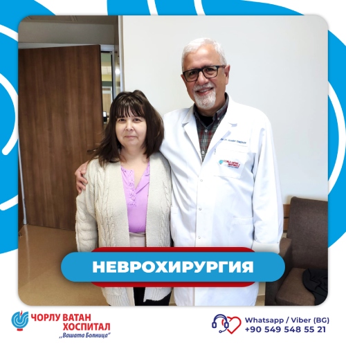
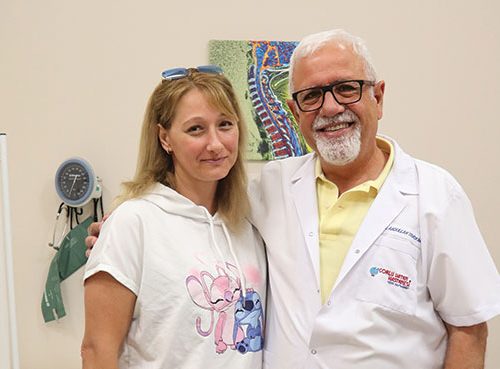
After more than two weeks of exhausting lower back pain radiating down the right leg and severe discomfort, our 45-year-old patient found relief at Corlu Vatan Private Hospital. Under the guidance of Dr. Abdullah Temizkan, a successful surgery was performed to remove a disc herniation in the lumbar segment of the spinal column.

The detailed physical examination by Dr. Abdullah Temizkan - neurosurgery specialist - revealed serious symptoms: muscle weakness, limited mobility, and a compressed nerve causing acute pain.
After an MRI confirming the diagnosis, the need for surgical intervention was discussed in detail, and the patient accepted. After the necessary preoperative preparation, Dr. Temizkan and his team proceeded to action. During the intervention, the herniated disc exerting pressure on the nerve roots was carefully removed. The result was almost instant pain relief and restoration of mobility.

The patient spent the night after the surgery under observation, and the very next day she was discharged in stable condition, with a smile and relief. She expressed her deep gratitude to Dr. Abdullah Temizkan and the entire team of Corlu Vatan Hospital for their care, attention, and professionalism.

Corlu Vatan Hospital thanks you for your trust and wishes our patient health, energy, and a new beginning - pain-free!

Corlu Vatan Private Hospital - "Your Hospital"

Yılan Yapısı 762862725
Successful Treatment of Cervical Disc Herniation and Spinal Stenosis

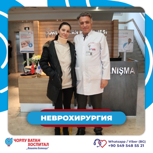
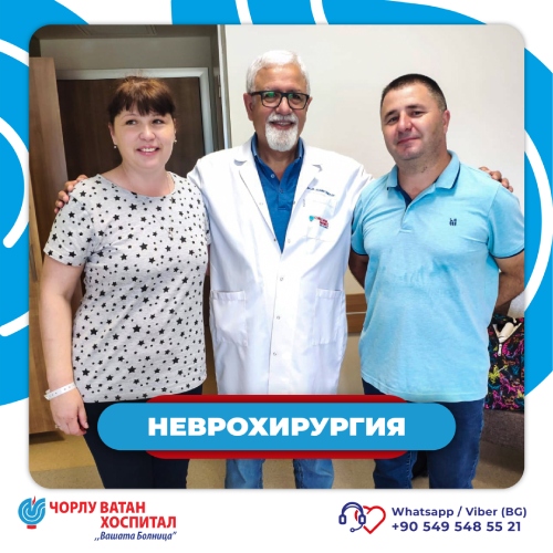
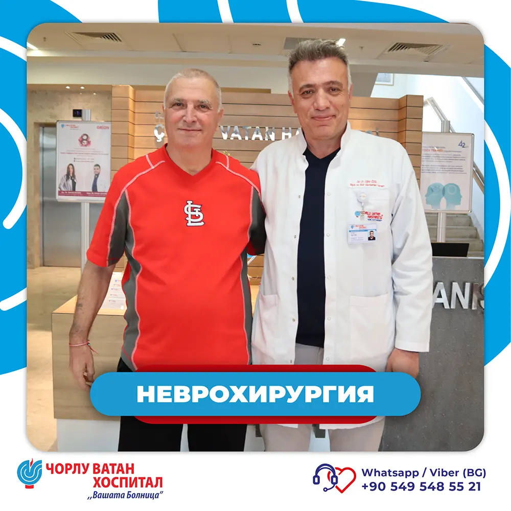
Our 43-year-old patient was admitted to Corlu Vatan Private Hospital with severe neck and right arm pain lasting more than a month.

After detailed examinations and tests conducted by Dr. Oguz Yozel, Neurosurgery Specialist, it was determined that the patient had loss of sensation in the cervical spine and compression of the nerve roots by damaged intervertebral discs.

During the surgery, the herniated discs were removed and replaced with an artificial cervical disc prosthesis, which restores mobility and reduces pressure on the nerves.

The procedure was successful, and the very next day the patient was discharged in good health. He expressed his gratitude to Dr. Oguz Yozel and the entire medical team.

The team of Corlu Vatan Hospital wishes him a speedy recovery and a healthy life!

Corlu Vatan Private Hospital - "Your Hospital"

Yılan Yapısı 2103274231
Active Again, Free Again!

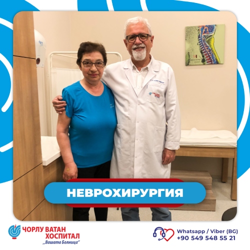
Lower back pain and numbness in the legs had been limiting the life of Mrs. Zdravka Todorova from Bulgaria for a long time. After a physical examination and tests, she was diagnosed with lumbar disc herniation. She chose to entrust her care to our neurosurgery specialist Dr. Abdullah Temizkan and the team at Corlu Vatan Hospital.

The surgery was successful, and today the patient once again enjoys freedom of movement and a life without pain.

Every person deserves a new beginning and a life without pain and limitations - we are here to help you.

If you suffer from lower back pain, numbness, or loss of strength in your legs - schedule a consultation appointment and take the first step toward a pain-free life!