


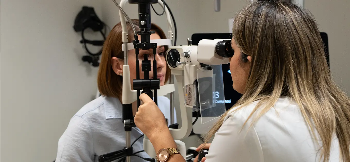
Thanks to the computerized eye health diagnostic systems we use in our Ophthalmology clinic, existing conditions in patients such as myopia, hyperopia, and astigmatism can be diagnosed, and appropriate glasses or contact lenses can be prescribed.

In patients with diabetes, improper regulation of blood sugar can lead to deterioration of blood vessels, which may result in retinal hemorrhages. If these hemorrhages are not treated in time, they can spread throughout the entire eye and cause permanent vision impairment. In our clinic, these hemorrhages can be detected at an early stage through fundus examination and application of special eye drops to dilate the pupil. Thanks to optical coherence tomography, the edema that may occur in the visual center is detected and can be treated with intravitreal injection. Additionally, with a retinal angiography device, vessels that are likely to bleed are identified and treated with a method called retinal laser without bleeding.
Macular degeneration (yellow spot disease) is one of the most important causes of poor vision, especially in elderly patients. With the help of optical coherence tomography and retinal angiography, this disease can be detected at its early stages, and if necessary, the progression of the disease can be prevented through treatment with intravitreal injections.
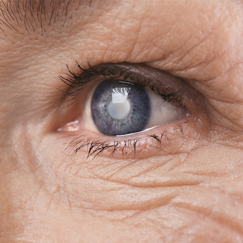
The cataractous lens is removed using the Phaco method (Phacoemulsification) and an artificial lens is placed in its position through a sutureless surgery.
With advancing age, cataract formation is common, which involves the loss of transparency of the eye's lens. Once a cataract has formed, it cannot be corrected by any means other than surgical intervention. In our Ophthalmology department, we apply the PHACO method to remove the cataractous lens, and an artificial lens is placed in the position of the removed natural lens of the eye.

Glaucoma is the second most common cause of vision impairment in the world after macular degeneration. Glaucoma is a disease that progresses insidiously without any symptoms, damaging the optic nerve and leading to permanent vision loss. The optic nerve damage caused by glaucoma cannot be reversed. Therefore, especially after the age of 40, patients with a family history of glaucoma or diabetes should have a routine comprehensive examination, even if they have no symptoms. Thanks to optical coherence tomography, optic nerve damage can be detected at a very early stage, and intraocular pressure can be controlled before the disease has progressed. Additionally, thanks to the special software of the tomography device used in our hospital, glaucoma progression can be monitored by comparing previous measurements during the patient's follow-up examinations.
Especially with aging, due to deterioration of the eyelid condition, the eyelashes may turn inward or the eyelids may turn outward from the eye. In both cases, burning, irritation, redness, and even tissue deterioration can occur in the eye, as the protective mechanism of the eyelids is compromised. In these cases, the eyelids can be restored to their previous position using special surgical techniques.
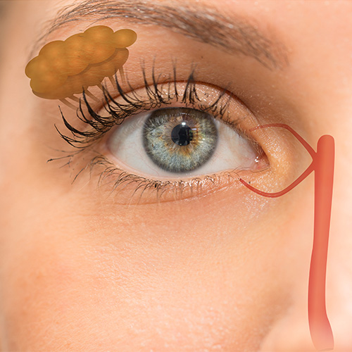
The tear duct starts from the lacrimal sac and passes toward the nose. A blockage in any part of this duct can be permanently corrected by attaching a temporary silicone tube to the area.
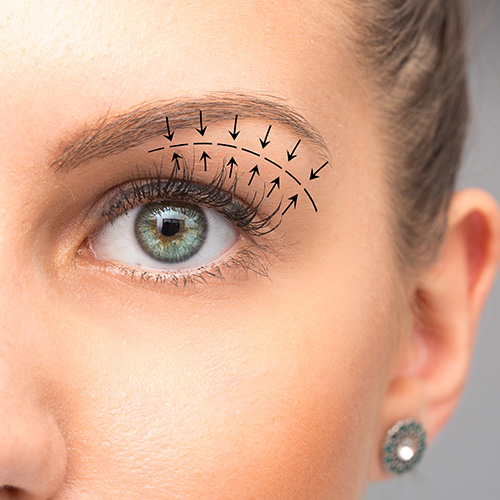
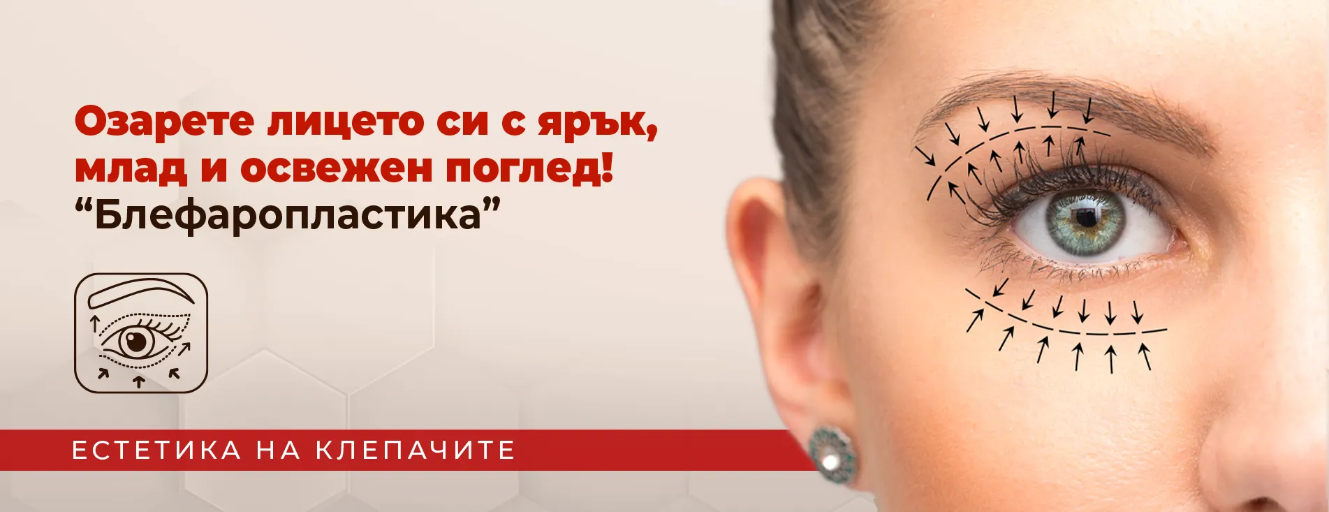
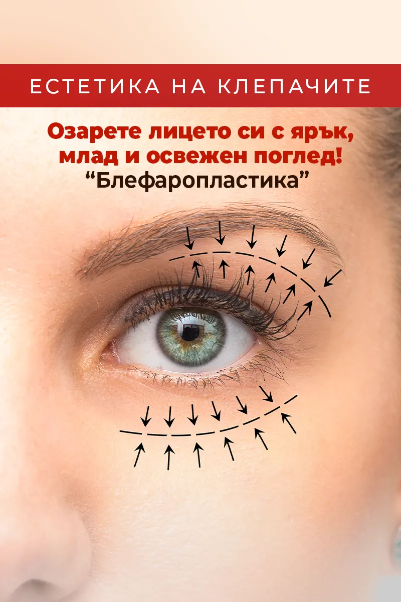
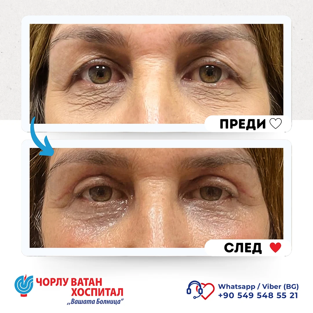
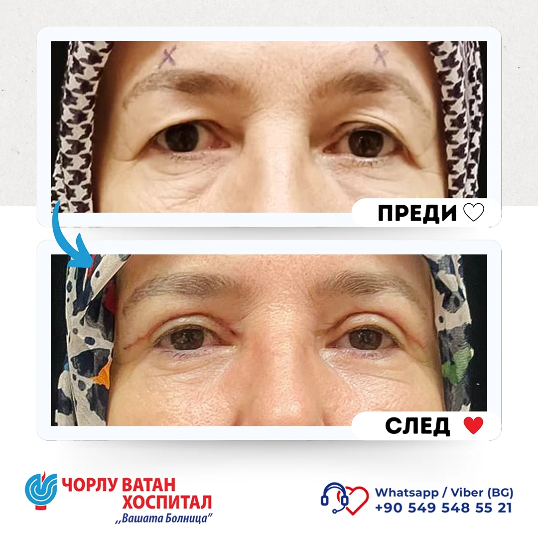
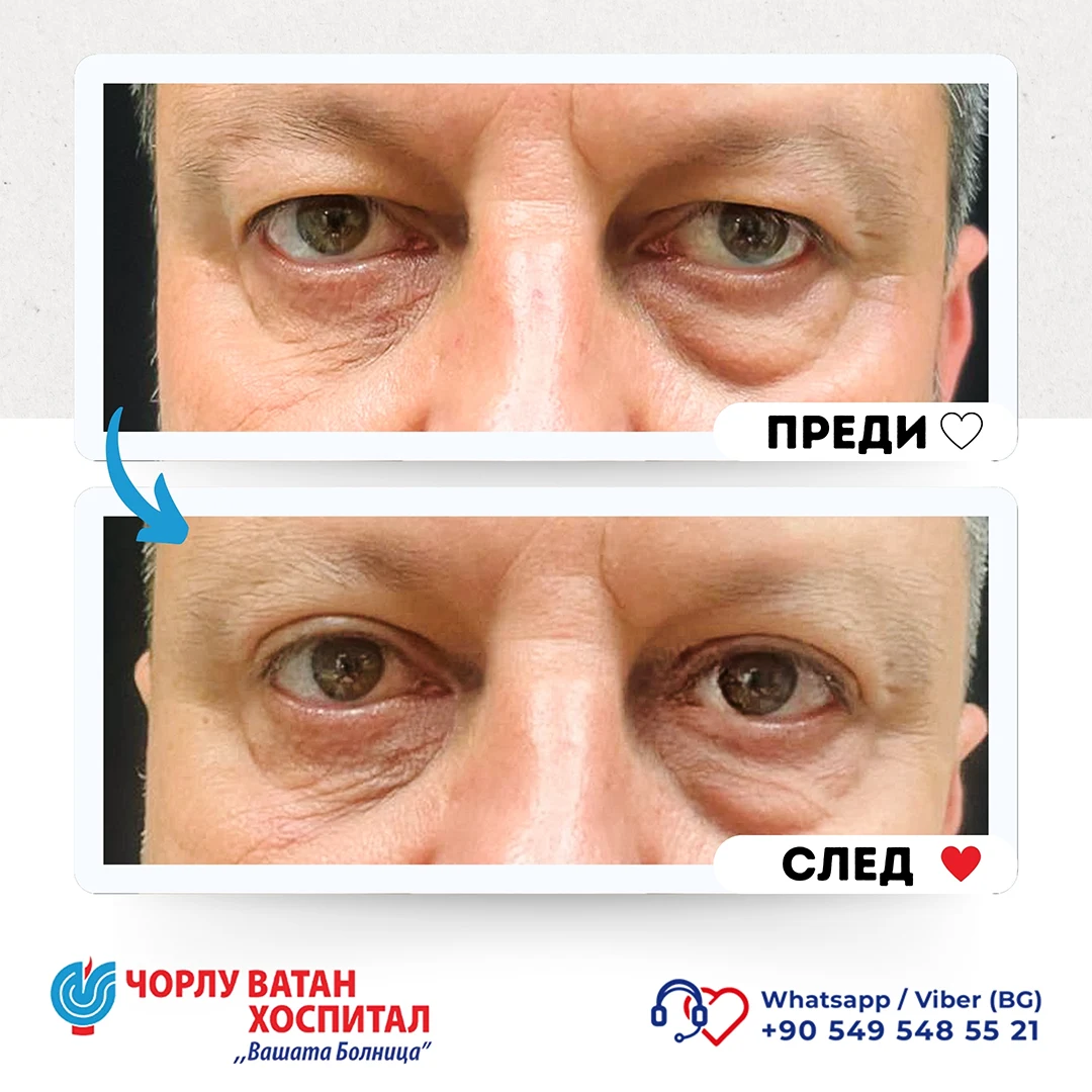
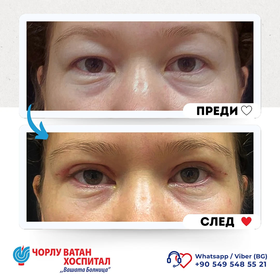
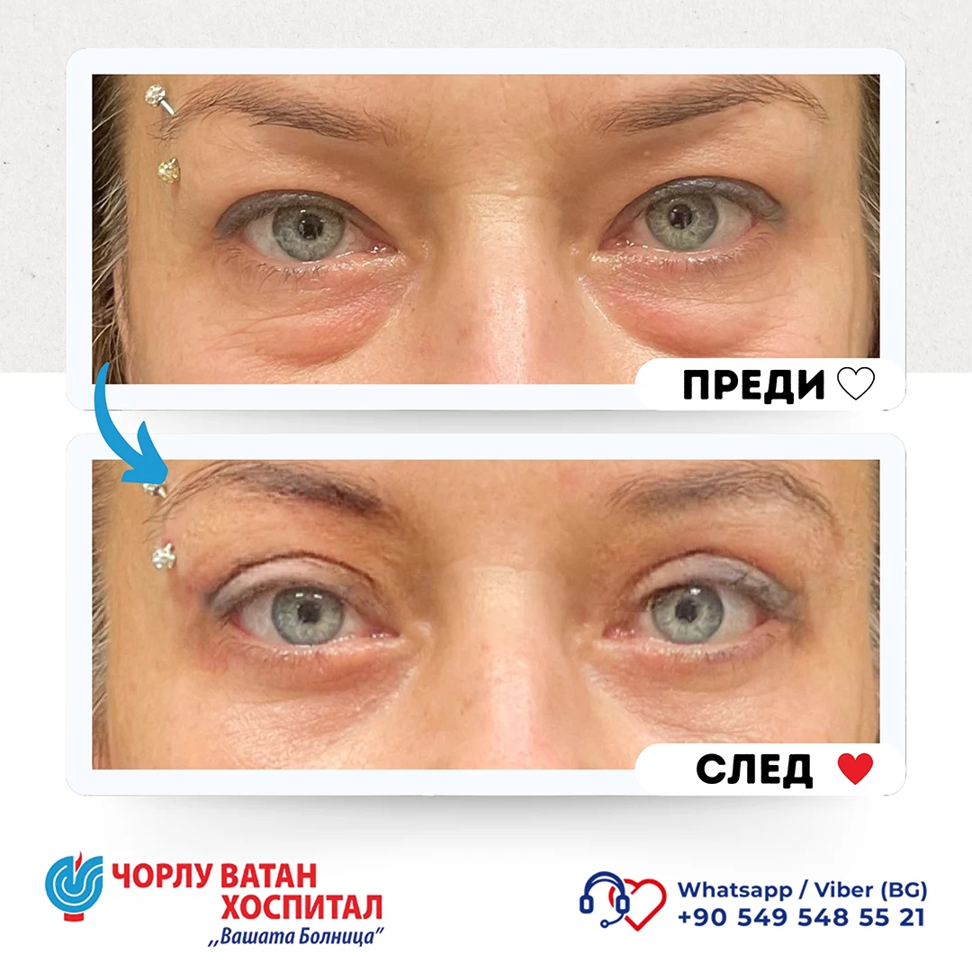
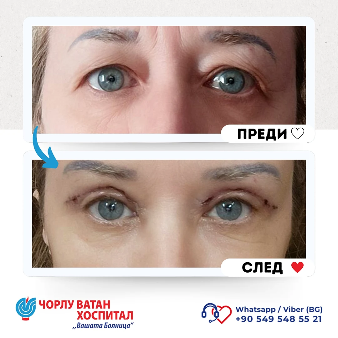
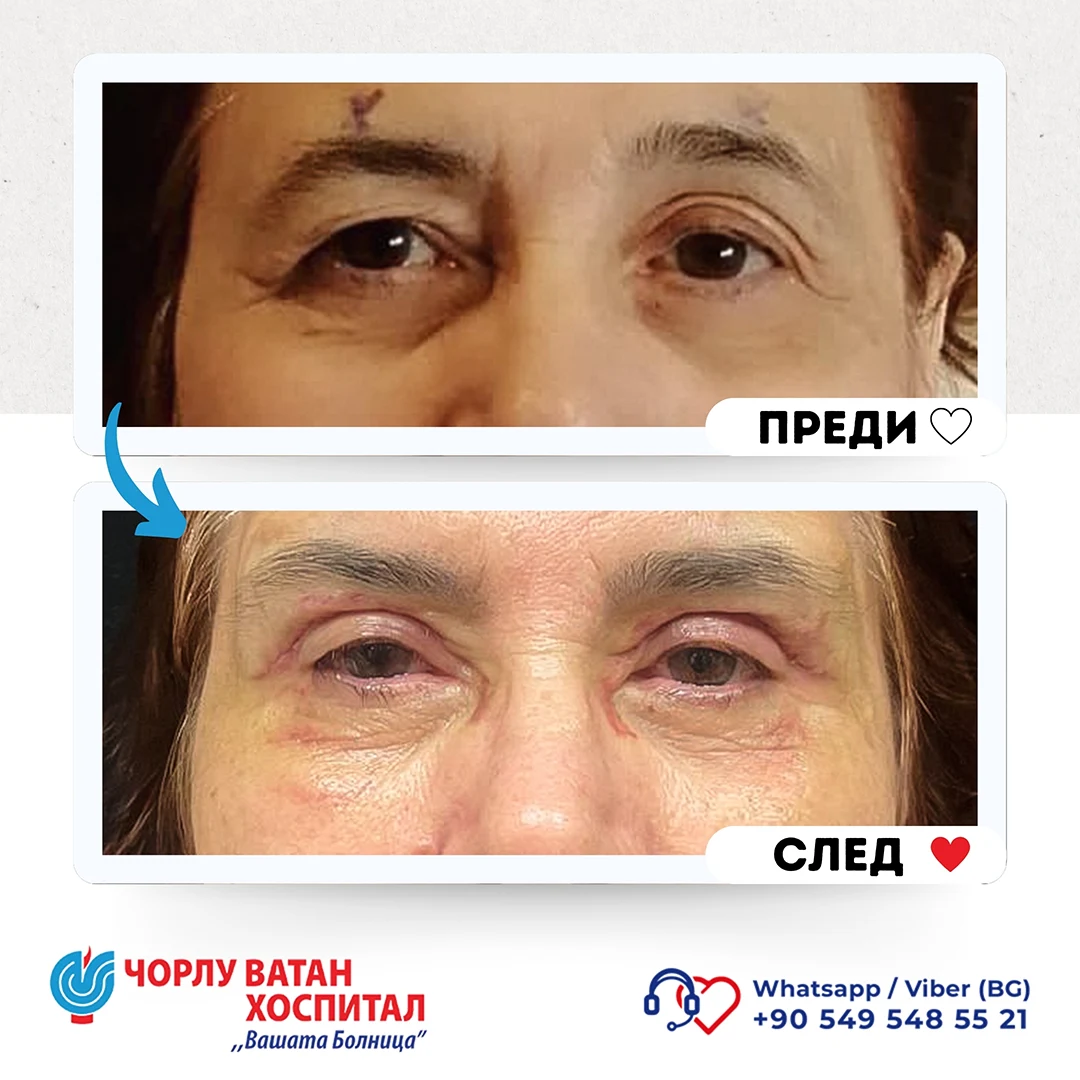
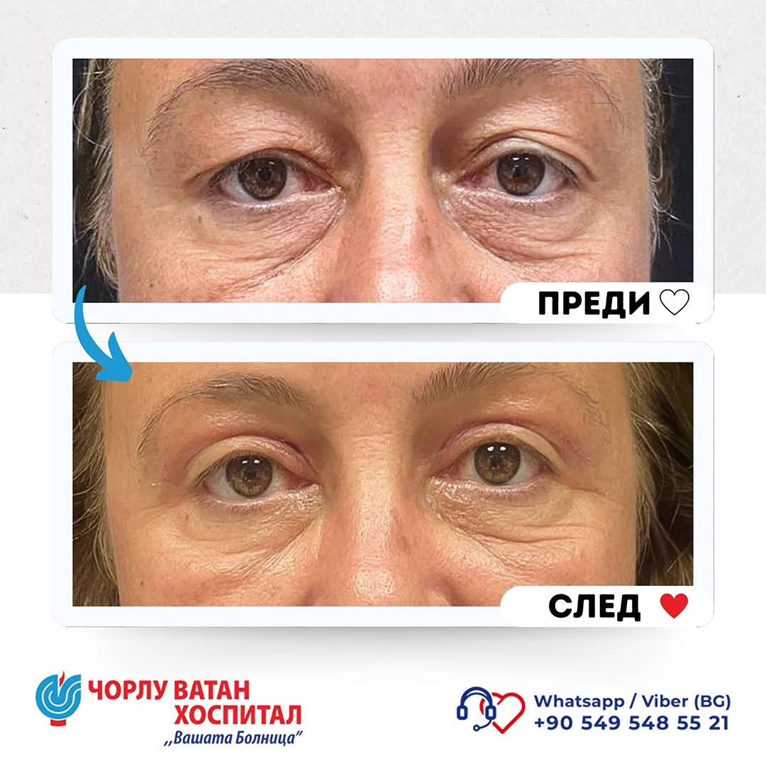
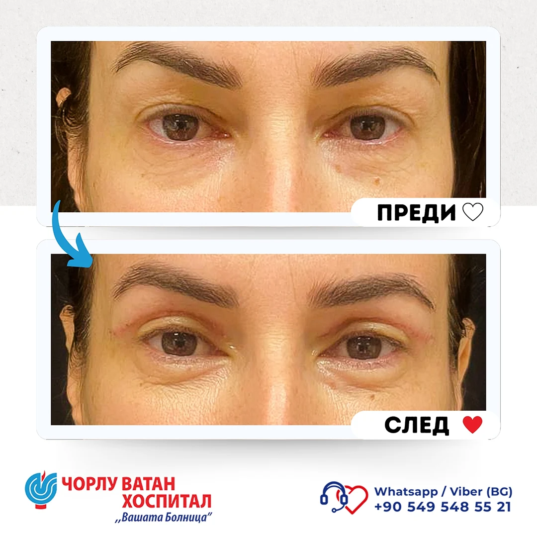
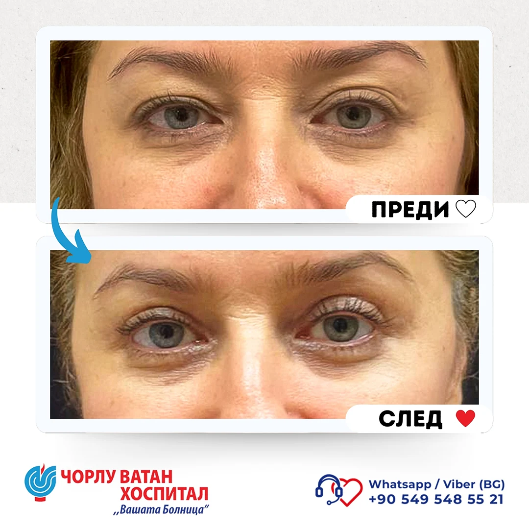
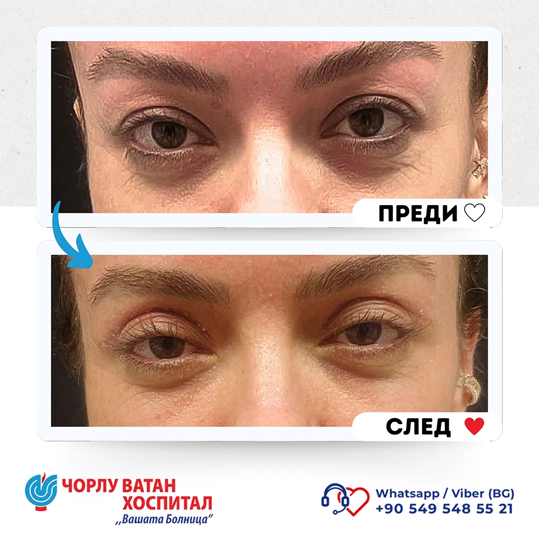
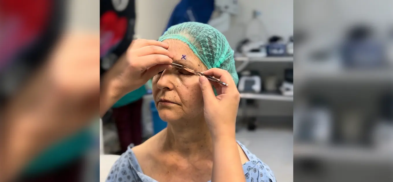
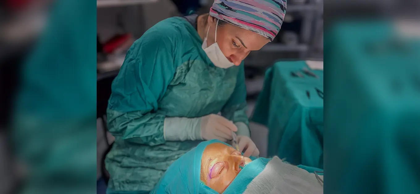
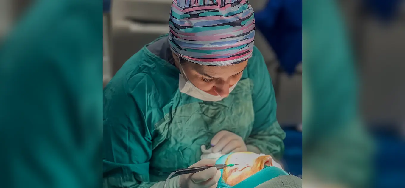
Upper eyelid blepharoplasty is a surgery performed when there is loose and excess skin and fatty deposits in the upper eyelid area, with the main cause being the aging process, and sometimes structural or pathological reasons. After a detailed physical examination and explanation, a surgical intervention plan is prepared. During the surgery, the incision line is planned to correspond to the upper eyelid crease, and during the intervention, excess muscle and fatty tissue are removed. Then, self-absorbing sutures are placed. In this surgery, scars are hidden in the eyelid crease so they are not visible. During the initial consultation with the patient, features such as drooping eyebrows and other possible problems in the periocular area are also evaluated, and options for their correction during the surgical intervention are discussed.
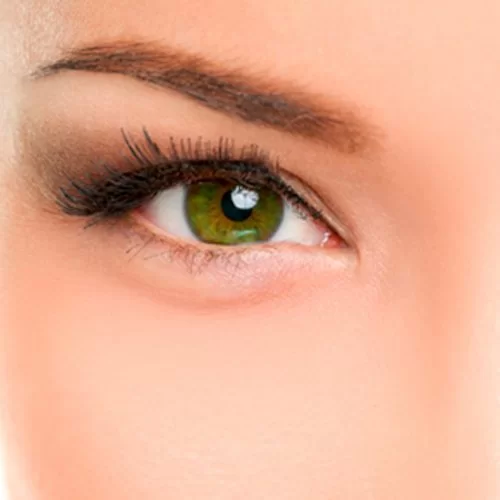
Almond eye surgery, which has become increasingly popular worldwide in recent years, gives the eyes a more youthful appearance by pulling the outer eye corners of the eyelids upward. This achieves a more refreshed and youthful look in the eye area, allowing patients to say goodbye to a tired facial expression. After evaluating the patient's condition, appropriate surgical planning can be made according to the desired eye shape. In our hospital, the procedure is performed using two methods.
In the first method, the outer corner of the eyelid is attached to the bone membrane with special permanent sutures using a surgical method.
The second method involves a special lifting technique using meso-threads. This is a method that uses threads that are absorbed over time. Together with the eyebrow, the eyelid edges are pulled upward toward the temple and secured with a special thread. This is not a surgical intervention and is the ideal method for patients who do not wish to undergo surgery. The average duration of the effect achieved by this method is 1-2 years. At the patient's request, after the effect of this method wears off, the same procedure can be repeated.
When deciding on almond eye surgery, proper evaluation of the patient's condition is very important. The best results are visible in patients where the outer corner of the eyelid is lower than the inner corner.
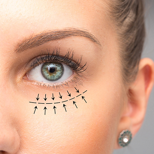
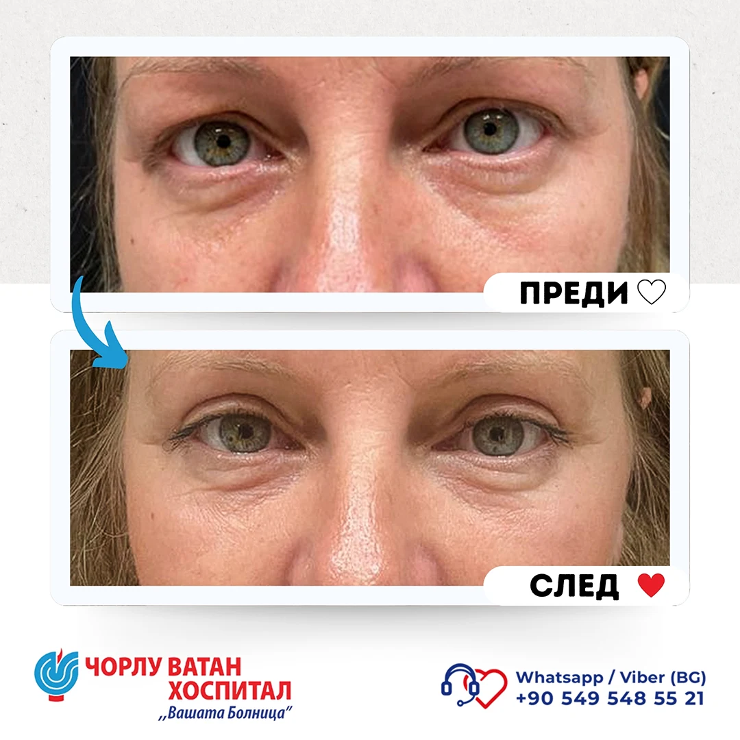
The skin around the eyes is the thinnest skin tissue in the body. For this reason, especially with age, the fat bags under the eyes become visible as the structures that hold the lower eyelid weaken. In transconjunctival lower eyelid surgery, these fat bags can be removed through a method where an incision is made from the inner side of the lower eyelid. This way, the appearance of the lower eyelid can be improved without a scar on the outer part of the eyelid. It is very important that the patient is suitable for this type of lower eyelid surgery. This surgery is not recommended for patients with a negative eye vector. After a detailed evaluation of the patient's condition, the most appropriate method for the patient is determined, and if the specific case is not suitable for this type of surgery, the method of Under-eye Lipolysis (mesotherapy with fat dissolving) is used.
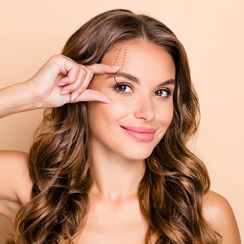
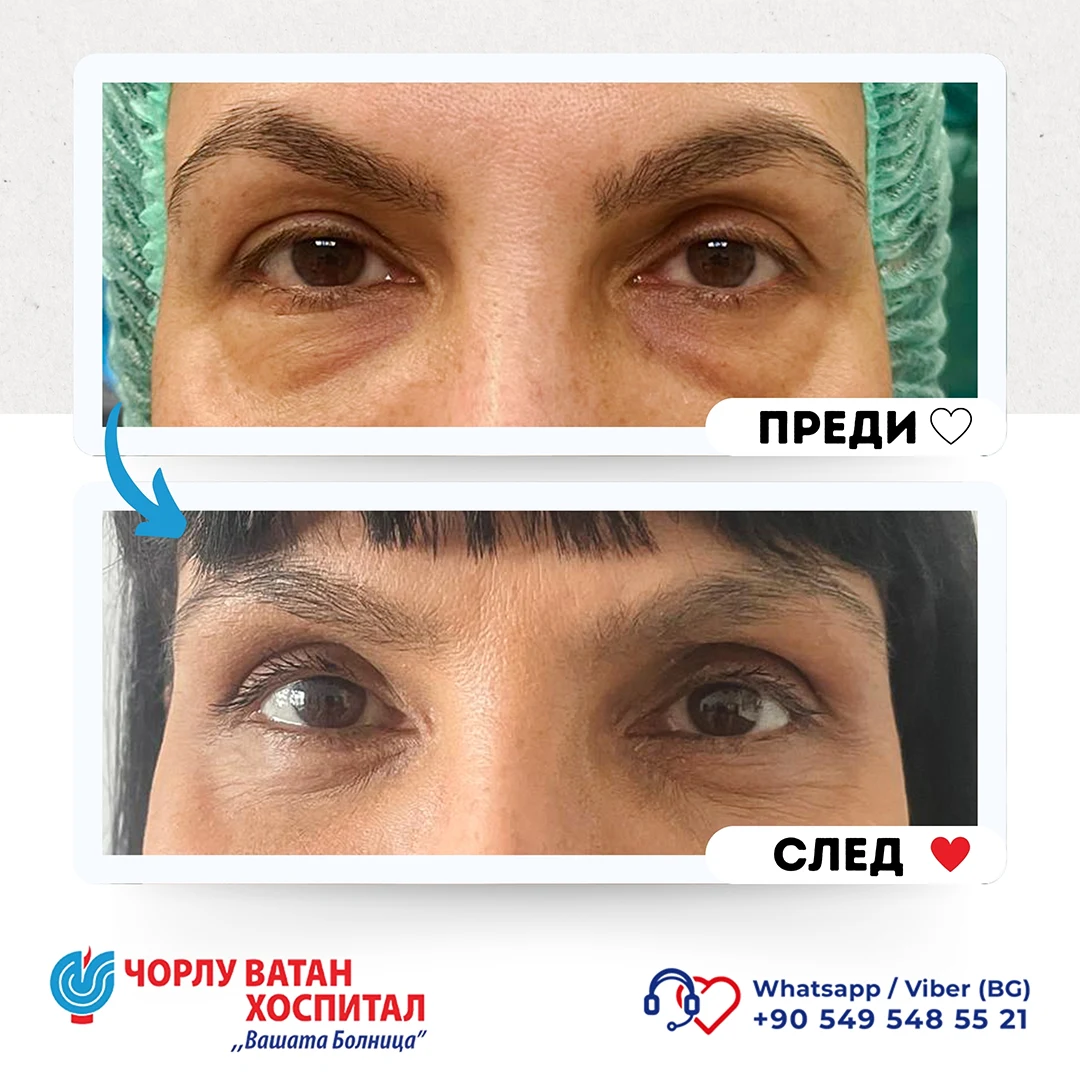
In patients where the lateral part of the eyebrow falls below the upper orbital bone, the eyebrow can be positioned higher with an incision from the inner side or just a small incision right above the eyebrow during upper eyelid surgery. A higher position of the lateral part of the eyebrow gives patients a younger and fresher appearance. In patients who do not wish to undergo surgical intervention, this part of the eyebrow can be lifted upward using absorbable meso-thread lifting methods.
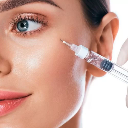
In our hospital, Botox (botulinum toxin) injection procedures are successfully performed in the forehead area and the eye area where "Crow's Feet" wrinkles are present. For patients with prominent under-eye grooves, periocular contour restoration with light filler is performed. Cheek area filling procedures are also applied to achieve lower eyelid tightening. For the treatment of dark circles and bruising in the under-eye area and to smooth wrinkles in this area, procedures such as mesotherapy are applied. For patients who have fat deposits in the under-eye area, if their case is not suitable or the patient does not wish to undergo surgical intervention, injection lipolysis procedures are successfully applied.
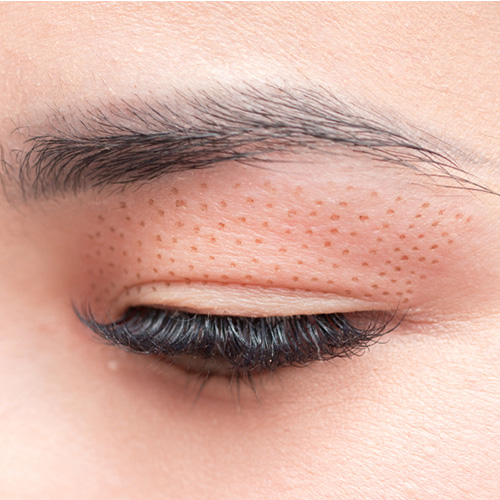
The fourth state of matter is called plasma. If a substance is supplied with energy when it is in a solid state, it first converts to a liquid and then to a gas. If we continue to supply energy to the substance while it is in a gaseous state, then plasma will be produced. This plasma energy can be used in healthcare to remove unwanted tissue or cellular formations from the surrounding environment without damaging the surrounding tissues.
Unwanted tissues on the surface are sublimated with the energy generated at the tip of the device application. Sublimation is the evaporation of cells called keratinocytes in the epidermis, i.e., the upper layer of the skin. The most important difference from laser and cautery is that there is no unwanted energy or heat flow to the surrounding environment and subcutaneous tissues. This way, the side effect profile is also very low. To perform these procedures, three different energy levels are used:
- Low energy consumption; eyelid procedures in blepharoplasty, skin spots, small warts, active acne, wrinkles, periumbilical stretch marks, and xanthelasma
- Medium energy; slightly larger warts, lesions, and deeper wrinkles
- High energy, on the other hand; is used for difficult-to-treat lesions, such as large lesions and keloids.
The procedure is a very good alternative for patients who have excess skin around the eyes but do not want to undergo surgery or do not need a surgical procedure. In such patients, it is called non-surgical blepharoplasty. It can be used to smooth the skin in patients with fine wrinkles under the eyes or in patients with excess skin after lower eyelid blepharoplasty. Especially in patients with fat deposits that cause festoon-like sagging from the lower eyelids to the cheeks, the results are very good when combined with lipolytic mesotherapy. It is very useful for removing lesions in patients with small sebaceous glands, skin folds, or warts around the eyes. In patients who have previously undergone eyelid surgery, in cases requiring touch-ups such as asymmetry, excess tissue, or poor scars, problems can be resolved without repeat surgery.
After applying a local anesthetic cream to the treatment area, disinfection of the area is performed. The procedure takes approximately 30-40 minutes. After the procedure, crusts form in the areas where the laser was applied. The crusts fall off within about 1 week. Underneath the crusts, new, healthy and fresh pink skin appears. It is very important to protect this area from ultraviolet rays (UV rays) for about 5 weeks. Swelling may occur for about 2-3 weeks. Usually (in most cases) one session is sufficient, but in patients with heavy eyelids, excess skin, and fat herniation, more than one session may be needed for optimal results.
The most important side effect is hyperpigmentation, which can occur if not protected from ultraviolet rays (UV rays). With good UV protection, such a problem does not arise.
The effect lasts on average about 2-3 years.


 Phone
Phone